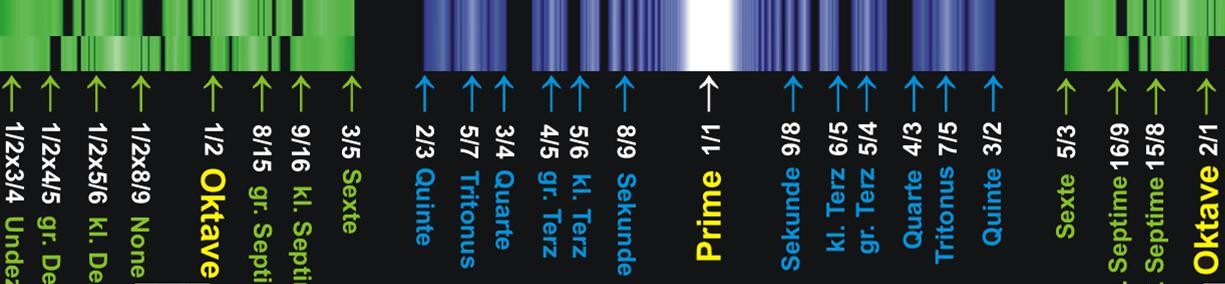
Magnetic Resonance Imaging
Proton nuclear magnetic resonance (NMR) detects the presence of hydrogens (protons) by subjecting them to a large magnetic field to partially polarize the nuclear spins, then exciting the spins with properly tuned radio frequency (RF) radiation, and then detecting weak radio frequency radiation from them as they „relax“ from this magnetic interaction. The frequency of this proton „signal“ is proportional to the magnetic field to which they are subjected during this relaxation process. In the medical application known as Magnetic Resonance Imaging (MRI), an image of a cross-section of tissue can be made by producing a well-calibrated magnetic field gradient across the tissue so that a certain value of magnetic field can be associated with a given location in the tissue. Since the proton signal frequency is proportional to that magnetic field, a given proton signal frequency can be assigned to a location in the tissue. This provides the information to map the tissue in terms of the protons present there. Since the proton density varies with the type of tissue, a certain amount of contrast is achieved to image the organs and other tissue variations in the subject tissue.
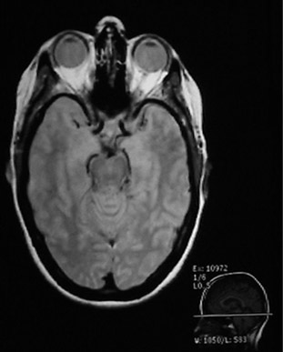 |
Since the MRI uses proton NMR, it images the concentration of protons. Many of those protons are the protons in water, so MRI is particularly well suited for the imaging of soft tissue, like the brain, eyes, and other soft tissue structures in the head as shown at left. The bone of the skull doesn’t have many protons, so it shows up dark. Also the sinus cavities image as a dark region.Bushong’s assessment is that about 80% of the body’s atoms are hydrogen atoms, so most parts of the body have an abundance of sources for the hydrogen NMR signals which make up the magnetic resonance image. |
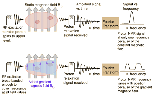
The schematic below may help visualize the imaging process. It is presumed that there are two regions of the sample which contain enough hydrogens to produce a strong NMR signal. The top sketch visualizes an NMR process with a constant magnetic field applied to the entire sample. The hydrogen spin-flip frequency is then the same for all parts of the sample. Once excited by the RF signal, the hydrogens will tend to return to their lower state in a process called „relaxation“ and will re-emit RF radiation at their Larmor frequency. This signal is detected as a function of time, and then is converted to signal strength as a function of frequency by means of a Fourier transformation. Since the protons in each of the active areas of the sample are subjected to the same magnetic field, they will produce the same frequency of radiation and the Fourier transform of the detected signal will have only one peak. This one peak demonstrates the presence of hydrogen atoms, but gives no information to locate them in the sample.
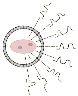 |
When a rotating field gradient is used, linear positioning information is collected along a number of different directions. That information can be combined to produce a two-dimensional map of the proton densities. The proton NMR signals are quite sensitive to differences in proton content that are characteristic of different kinds of tissue. Even though the spatial resolution of MRI is not as great as a conventional x-ray film, its contrast resolution is much better for tissue. Rapid scanning and computer reconstruction give well-resolved images of organs. |