Magnetic Resonance Imaging
Proton nuclear magnetic resonance (NMR) detects the presence of hydrogens (protons) by subjecting them to a large magnetic field to partially polarize the nuclear spins, then exciting the spins with properly tuned radio frequency (RF) radiation, and then detecting weak radio frequency radiation from them as they „relax“ from this magnetic interaction. The frequency of this proton „signal“ is proportional to the magnetic field to which they are subjected during this relaxation process. In the medical application known as Magnetic Resonance Imaging (MRI), an image of a cross-section of tissue can be made by producing a well-calibrated magnetic field gradient across the tissue so that a certain value of magnetic field can be associated with a given location in the tissue. Since the proton signal frequency is proportional to that magnetic field, a given proton signal frequency can be assigned to a location in the tissue. This provides the information to map the tissue in terms of the protons present there. Since the proton density varies with the type of tissue, a certain amount of contrast is achieved to image the organs and other tissue variations in the subject tissue.
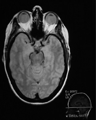 |
Since the MRI uses proton NMR, it images the concentration of protons. Many of those protons are the protons in water, so MRI is particularly well suited for the imaging of soft tissue, like the brain, eyes, and other soft tissue structures in the head as shown at left. The bone of the skull doesn’t have many protons, so it shows up dark. Also the sinus cavities image as a dark region.Bushong’s assessment is that about 80% of the body’s atoms are hydrogen atoms, so most parts of the body have an abundance of sources for the hydrogen NMR signals which make up the magnetic resonance image. |


 Die Musik des Universums ist eine große Harmonie. Von den feinsten Schwingungen der Atome bis zu den groben Bewegungen unserer Planeten ist alles miteinander kombiniert.